Ayahuasca Stimulates the Birth of New Brain Cells
For a long time, scientists believed that no new neurons are born in the brains of adults, meaning that when our existing brain cells become damaged or die, they are not replaced. However, it was later discovered that neurogenesis – meaning the creation of new neurons – does in fact in occur in the hippocampus, a brain region associated with memory. Unfortunately, the rate of neurogenesis is not always sufficient to replace all of our damaged neurons as we age, which is why many people suffer from dementia and other age-related cognitive deficiencies. Fortunately, a study conducted by the Beckley/Sant Pau Research Programme, and published in the journal Scientific Reports, reveals that certain compounds present in the psychedelic Amazonian brew ayahuasca actually stimulate the birth of new neurons.
Researchers placed harmine and tetrahydroharmine – the most prevalent alkaloids in ayahuasca – in a petri dish with hippocampal stem cells, and found that this greatly increased the rate at which these cells developed into fully mature neurons. The results of this study were first presented at the Interdisciplinary Conference on Psychedelics Research in 2016, and represent the first evidence that components of ayahuasca have neurogenic properties, thereby opening up a wealth of possibilities for future research.
We are currently conducting additional experiments to discern the magnitude of the observed effects, as well as undertaking studies on live animals. The replication of the present findings in vivo would represent a major breakthrough in mental healthcare, with potential applications ranging from treating neurodegenerative and psychiatric disorders to redressing brain damage associated with stroke or trauma.
Jordi Riba of the Beckley/Sant Pau Research Programme explains the latest findings, illustrating them with the beautiful images below
What you are seeing is a “static picture” taken after several days of treatment of the stem cells with the different compounds. No neurons were present prior to the three different tretments: a) saline (water+salt); b) harmine; and c) tetrahydroharmine
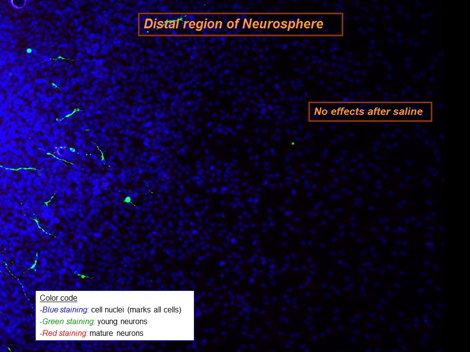
The first image is the control, when only salty water (saline) added to the cell cultures. The nuclei of the stem cells can be seen in blue.These stem cells have been treated with saline for several days and only a few have developed into young neurons (the few green sports in the image).
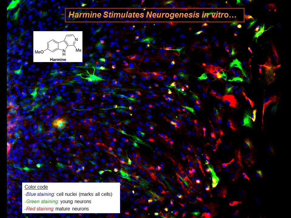
The second image shows the results after several days of treatment with harmine: blue is still present because it’s a marker of cell nuclei, and all cells have nuclei (stem cells and neurons). The green spots are the young neurons marked using Tuj1 staining (this staining is specific for “neuron-specific class III beta-tubulin) present in recently created neurons. The red spots show more mature neurons. The staining marks the “microtubule-associated protein 2 (MAP-2). Its presence increases during neuron development.
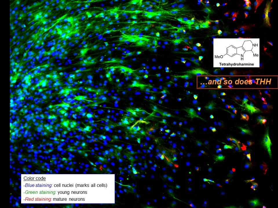
The third image shows the results obtained after several days of treatment with tetrahydroharmine. The meaning of the colors is the same.
Words: Jordi Riba
Head of the Human Neuropsychopharmacology Research Group at Sant Pau Hospital in Barcelona and a co-director with Amanda Feilding, of the Beckley/Sant Pau Research Programme
Podcast
- All
Links
- All
Support
- All
BIPRP
- All
Science Talk
- All
Amanda's Talks
- All
- Video Talk
- Featured
- 2016 Onwards
- 2011-2015
- 2010 and Earlier
- Science Talk
- Policy Talk
One-pager
- All
Music
- All
Amanda Feilding
- All
Events
- All
Highlights
- All
Psilocybin for Depression
- All
Current
- All
Category
- All
- Science
- Policy
- Culture
Substance/Method
- All
- Opiates
- Novel Psychoactive Substances
- Meditation
- Trepanation
- LSD
- Psilocybin
- Cannabis/cannabinoids
- Ayahuasca/DMT
- Coca/Cocaine
- MDMA
Collaboration
- All
- Beckley/Brazil Research Programme
- Beckley/Maastricht Research Programme
- Exeter University
- ICEERS
- Beckley/Sant Pau Research Programme
- University College London
- New York University
- Cardiff University
- Madrid Computense University
- Ethnobotanicals Research Programme
- Freiburg University
- Medical Office for Psychiatry and Psychotherapy, Solothurn
- Beckley/Sechenov Institute Research programme
- Hannover Medical School
- Beckley/Imperial Research Programme
- King's College London
- Johns Hopkins University
Clinical Application
- All
- Depression
- Addictions
- Anxiety
- Psychosis
- PTSD
- Cancer
- Cluster Headaches
Policy Focus
- All
- Policy Reports
- Advisory Work
- Seminar Series
- Advocacy/Campaigns
Type of publication
- All
- Original research
- Report
- Review
- Opinion/Correspondence
- Book
- Book chapter
- Conference abstract
- Petition/campaign
Search type